Science, Corporate Abigail Chard 14/12/2023 Science, Corporate Abigail Chard 14/12/2023 Winners of £50K Franklin research residencies announced Read More Science Abigail Chard 09/11/2023 Science Abigail Chard 09/11/2023 Left-handers aren’t better spatially, gaming research proves Read More Charity, Corporate, Engineering, Environment, Health & Medicine, Science, Social Sciences Richard Ashby 08/09/2023 Charity, Corporate, Engineering, Environment, Health & Medicine, Science, Social Sciences Richard Ashby 08/09/2023 Benchtop NMR spectroscopy can accurately analyse pyrolysis oils Read More Health & Medicine, Science Richard Ashby 15/06/2023 Health & Medicine, Science Richard Ashby 15/06/2023 World’s first picture of the molecular machinery that makes cilia beat Read More Engineering, Environment, Science Richard Ashby 30/03/2023 Engineering, Environment, Science Richard Ashby 30/03/2023 New glass sensors could make commercial nuclear fusion viable Read More Health & Medicine, Science Richard Ashby 11/03/2023 Health & Medicine, Science Richard Ashby 11/03/2023 Urine gene test can predict bladder cancer years before diagnosis Read More Health & Medicine, Science Richard Ashby 23/01/2023 Health & Medicine, Science Richard Ashby 23/01/2023 Split-second of evolutionary mutation could have led to mammals, says UCL researcher Read More Science Richard Ashby 20/12/2022 Science Richard Ashby 20/12/2022 Signal processing algorithms improved turbulence in free-space optic tests Read More
Science, Corporate Abigail Chard 14/12/2023 Science, Corporate Abigail Chard 14/12/2023 Winners of £50K Franklin research residencies announced Read More
Science Abigail Chard 09/11/2023 Science Abigail Chard 09/11/2023 Left-handers aren’t better spatially, gaming research proves Read More
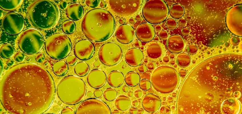
Charity, Corporate, Engineering, Environment, Health & Medicine, Science, Social Sciences Richard Ashby 08/09/2023 Charity, Corporate, Engineering, Environment, Health & Medicine, Science, Social Sciences Richard Ashby 08/09/2023 Benchtop NMR spectroscopy can accurately analyse pyrolysis oils Read More
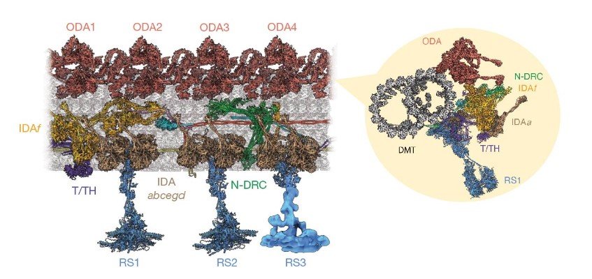
Health & Medicine, Science Richard Ashby 15/06/2023 Health & Medicine, Science Richard Ashby 15/06/2023 World’s first picture of the molecular machinery that makes cilia beat Read More
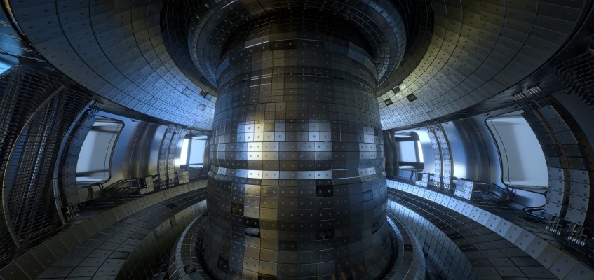
Engineering, Environment, Science Richard Ashby 30/03/2023 Engineering, Environment, Science Richard Ashby 30/03/2023 New glass sensors could make commercial nuclear fusion viable Read More
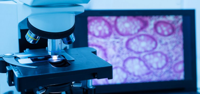
Health & Medicine, Science Richard Ashby 11/03/2023 Health & Medicine, Science Richard Ashby 11/03/2023 Urine gene test can predict bladder cancer years before diagnosis Read More
Health & Medicine, Science Richard Ashby 23/01/2023 Health & Medicine, Science Richard Ashby 23/01/2023 Split-second of evolutionary mutation could have led to mammals, says UCL researcher Read More
Science Richard Ashby 20/12/2022 Science Richard Ashby 20/12/2022 Signal processing algorithms improved turbulence in free-space optic tests Read More